
Executive Summary
A dog ear infection that returns within weeks or months of treatment is one of the most common frustrations in veterinary practice. Research shows the average time to relapse is just 3.6 months, and the reason is almost never the infection itself. Undiagnosed allergy, undertreated middle-ear disease, and growing antibiotic resistance in South African dog populations all drive the cycle. This article explains what is actually happening, and what needs to change to stop it.
The Recurring Ear Infection Cycle Most Owners Miss
A dog ear infection that clears up and then returns is not the same infection twice. It is a sign that the underlying cause was never identified or addressed. Veterinary researchers describe ear disease in dogs as a layered syndrome, not a single condition. A University of Pretoria clinical review frames it precisely this way: predisposing factors create a vulnerable canal, a primary cause triggers the initial inflammation, secondary organisms move in and infect the tissue, and perpetuating factors keep the environment hostile enough for the problem to survive treatment.
Most standard treatment courses target only the secondary infection. The ear drops clear the bacteria or yeast. The dog improves. Then, weeks later, the same warm, inflamed, allergy-affected canal allows the same organisms to recolonise, and the cycle starts again. Until each layer of that framework gets assessed and treated, recurrence is essentially guaranteed.
Allergy: The Root Cause of Canine Ear Disease
Canine ear disease overwhelmingly traces back to allergy. A clinical study of 100 dogs presenting with ear infections identified allergy as the primary cause in 43% of cases, making it the single largest driver by a significant margin. At specialist referral practices, up to 75% of all ear infection cases have an underlying allergic cause.
Two forms of allergy account for most of these cases. Canine atopic dermatitis is a hypersensitivity reaction to environmental allergens such as dust mites, grass pollen and mould spores. Cutaneous adverse food reactions involve an immune response to a specific protein in the diet. Both produce inflammation in the ear canal that changes the local environment, disrupting the normal skin barrier and encouraging bacterial and yeast populations to grow beyond their usual levels.
The ear is often the first place allergy shows up in a dog. Research notes that in roughly 43% of dogs with atopic dermatitis, the owner identified the ear as the initial site of the problem. Breed susceptibility runs strong through Labradors, Cocker Spaniels, West Highland White Terriers, German Shepherds and Boxers. Drop-eared breeds face additional structural risk, since reduced airflow in the canal traps moisture and warmth, compounding whatever the allergy has already started.
Treating a dog ear infection without investigating and managing the allergy leaves the primary cause fully intact. The canal stays inflamed between treatment courses. Conditions remain ideal for the next episode. This is why so many dogs cycle through antibiotics repeatedly without ever achieving lasting resolution of their ear problems.
The Recurring Ear Infection Cycle
🌿
Allergy Flares
The primary cause, atopy or food allergy, inflames the canal lining and disrupts the skin barrier. Bacteria and yeast begin to multiply.
⚠️
Infection Sets In
Secondary bacterial or yeast infection develops. Treatment clears the organisms, but the inflamed, allergy-affected canal remains. Average relapse: 3.6 months.
🔁
Cycle Repeats
With the primary cause unaddressed, the canal stays vulnerable. Each episode can introduce more resistant bacteria and deepen structural damage.
How Bacteria Turn a Dog Ear Infection Chronic
Secondary bacterial infection drives most of the pain, discharge and odour that owners associate with a dog ear infection. Two organisms dominate the picture. Staphylococcus pseudintermedius is the most common culprit in early and moderate infections. In chronic, difficult-to-treat cases, Pseudomonas aeruginosa takes over. It is an environmental organism that produces a protective biofilm inside the canal, physically shielding itself from both ear drops and the immune system.
Repeated treatment cycles without resolving the primary cause do something else: they select for resistant strains. Each incomplete course eliminates the most susceptible bacteria and leaves the resistant ones behind. Over time, the population in the canal shifts toward organisms that standard antibiotics cannot touch.
South Africa’s Resistance Problem
South African data from Onderstepoort makes this concrete. A study published in the Journal of the South African Veterinary Association in 2022, drawing isolates from five provinces, found that 85.9% of Staphylococcus pseudintermedius samples from canine skin and ear infections carried the mecA gene, the genetic marker for methicillin resistance. That resistance extends across all penicillin-type antibiotics. Resistance to enrofloxacin ran at 65% among those same isolates. Doxycycline and clindamycin fared only slightly better.
The Pseudomonas picture is worse still. A separate Onderstepoort study analysed 155 isolates from a single veterinary academic hospital and found that 92% were multidrug-resistant. Previous hospitalisation, a history of pruritus, and prior antibiotic treatment failure were each identified as significant risk factors for carrying a resistant strain. These are not fringe cases. They represent a large portion of the dogs presenting with recurring canine ear disease at veterinary practices across the country.
When Ear Canal Infection Spreads to the Middle Ear
Chronic ear canal infection creates a second, frequently missed complication. When infection and inflammation persist long enough, they degrade the eardrum and allow bacteria to cross into the middle ear. Research shows this complication occurs in 50–80% of chronic ear infection cases. The problem is that it often goes undetected, because the eardrum can appear intact even when the middle ear is already infected and inflamed behind it.
Middle-ear involvement changes the clinical picture significantly. Beyond the usual ear discomfort, a dog may show pain when opening its mouth, a head tilt, balance problems, or facial drooping on the affected side. It also changes the treatment requirements entirely. Topical ear drops do not reach the middle ear. Systemic antibiotics, guided by culture results and maintained for four to six weeks, become necessary. Failing to recognise middle-ear involvement is one of the most common reasons a dog ear infection appears to resist all treatment.
Why Standard Courses Fail Chronic Ear Problems
Standard antibiotic courses fail chronic ear problems for two connected reasons. The first is the resistance data already described. Prescribing a broad-spectrum antibiotic without first identifying the organism through cytology and culture means there is a very high probability the drug will not match the bacteria present. In South Africa, that probability runs at 85.9% for staphylococcal infections and climbs even higher for Pseudomonas.
The second reason is biofilm. Pseudomonas aeruginosa produces a dense extracellular matrix around itself inside the ear canal. Standard antibiotic ear drops cannot penetrate this structure at normal concentrations. Tris-EDTA, a chelating solution that strips divalent cations from the outer membrane of Gram-negative bacteria, disrupts biofilm and makes the canal accessible to antibiotics again. Instilling it and waiting 20–30 minutes before applying the antibiotic drops is standard practice in resistant Pseudomonas cases. Without this step, drops applied directly to a biofilm-covered canal achieve very little.
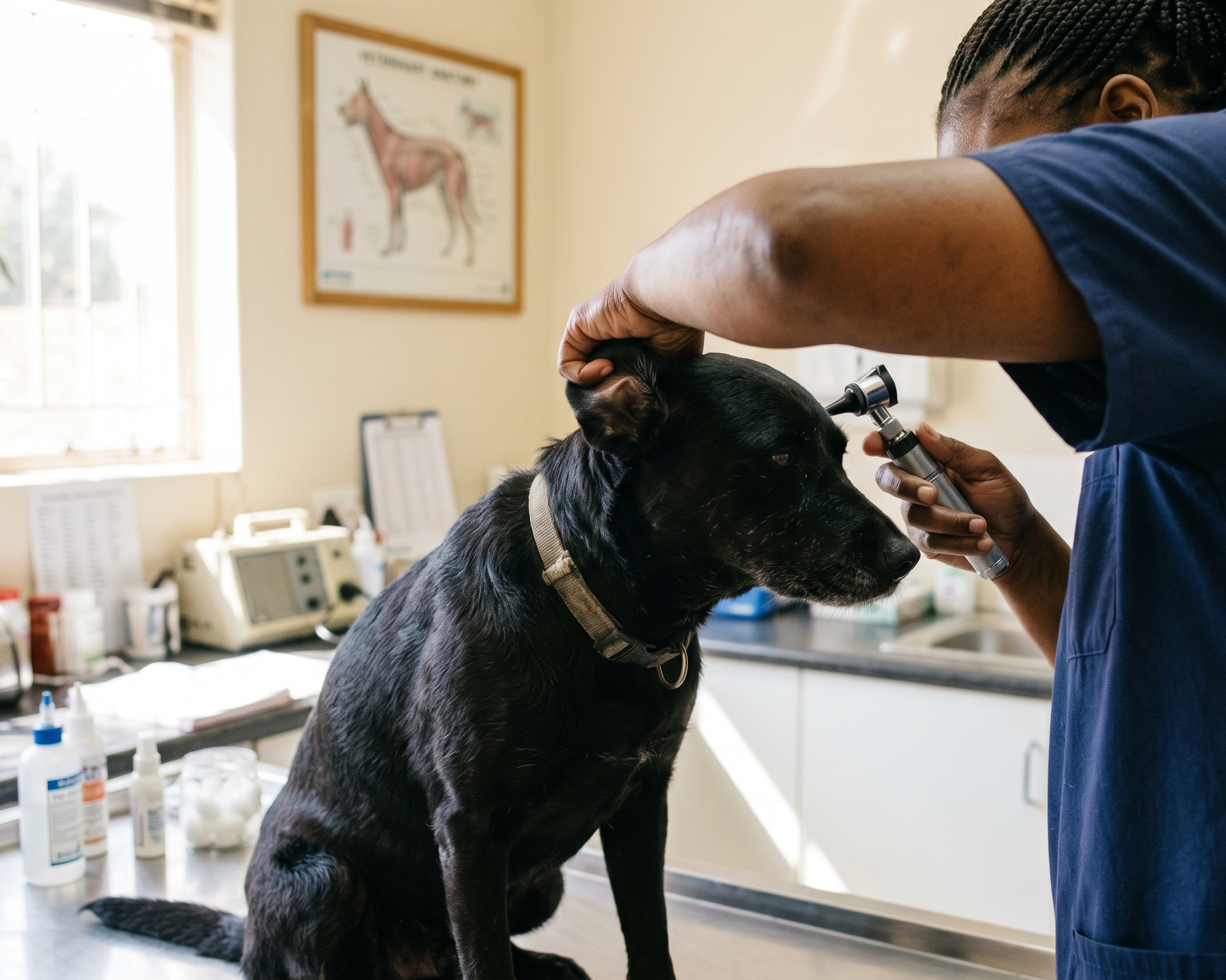
What a Correct Diagnosis Actually Looks Like
Cytology sits at the centre of every competent ear assessment. A sample from the canal, stained and examined under a microscope, tells the clinician which organisms are present and in what form. Cocci-shaped bacteria point toward Staphylococcus. Rod-shaped bacteria indicate Gram-negative organisms such as Pseudomonas, which should immediately trigger culture and sensitivity testing rather than empirical prescribing. Yeast cells in their characteristic budding form confirm a fungal component. Each finding leads to a different treatment path.
Beyond cytology, a thorough assessment of any recurring dog ear infection includes examination of the eardrum’s integrity, evaluation of the canal structure for thickening or calcification, and a full clinical history covering the pattern of recurrence, previous treatments and their outcomes, and any concurrent skin or digestive signs that might point toward allergy. CT or MRI imaging is the most reliable method for detecting middle-ear involvement when it is suspected and cannot be confirmed by otoscopy alone.
Breaking the Dog Ear Inflammation Cycle
Resolving dog ear inflammation on a lasting basis requires working through the problem in sequence. Clearing the active infection comes first, using targeted topical therapy matched to the cytology result. Thorough ear cleaning before applying drops matters: discharge and debris reduce the contact between medication and tissue, and saline flushing is safe even when the eardrum is compromised.
The second step is the one most often skipped. Identifying and managing the primary cause, which is almost always the allergy, determines whether the dog remains free of infection or relapses within months. A dietary elimination trial, run strictly for eight to twelve weeks with a novel or hydrolysed protein diet, rules out food allergy. Atopic dermatitis requires its own investigation and long-term management, whether through allergen-specific immunotherapy, anti-itch medication, or both.
Where middle-ear disease has developed, systemic antibiotics at appropriate doses for a full four-to-six-week course are necessary. Cases that have progressed to irreversible structural damage, with a calcified canal and entrenched infection that cannot be cleared medically, reach a point where surgical removal of the canal becomes the only remaining option. That outcome is preventable in most dogs, but only through earlier, more thorough investigation of why the first infection kept returning.
Sources
